Diagnosis first, then treatment
The lesions described on this page are not a uniform group. Some of them are a developmental vascular defect present from birth, some develop with age, and a few can look similar to lesions requiring a different approach.
This is why qualification is carried out by a doctor, and for lesions that are atypical, fast growing or bleeding, the first step is diagnostics, not removal. We support the assessment with digital dermatoscopy, but this aids the decision, it does not provide a diagnosis.
On this page
Three types of lesion that are easy to confuse
This distinction has practical significance, because it determines whether the lesion disappears after one treatment or requires a course and monitoring for years.
Dilated vessel, also known as telangiectasia
A single vessel that has permanently widened and become visible through the epidermis. Thin, running in a linear or star shaped pattern. It develops during life, most often under the influence of sun exposure, hormones or predisposition.
A closed vessel is absorbed by the body and does not return. New ones can continue to form.
Haemangioma
A lesion made up of overgrown vessels, forming a raised or flat structure. Ruby haemangiomas, small red dots, appear with age and are very common. Flat and deeper haemangiomas have a greater volume and reach further into the skin.
Removable, but the number of sessions depends on the volume and depth of the lesion.
Vascular malformation
A developmental vascular defect, present from birth, although not always visible straight away. It is not overgrowth but an abnormal structure of the vascular network. This group includes port wine stains and lesions such as venous lakes.
We do not treat this group of lesions. We include it so that it is possible to recognise that the problem requires a different centre.
If the lesion is a single thin vessel or diffuse facial redness, the right page is vessel closure and redness reduction. There we describe superficial lesions, here more complex ones.
Lesions we treat
Below are the lesions covered by laser therapy at our practice, with notes on what determines the price and the number of sessions.
Cherry angiomas
Small, raised red lesions, most often on the trunk and arms, appearing with age. Usually multiple. A benign lesion, removed for aesthetic reasons.
Per single lesion
Flat haemangiomas
Lesions with a larger surface area, flat or slightly raised, with a colour ranging from red to bluish. They usually require a course of treatments.
By surface area
Deeper and mixed haemangiomas
Lesions with a greater volume, involving vessels located deeper in the skin. They require a longer wavelength and higher energy, and often several sessions.
By surface area
Haemangioma on the vermilion of the lip
An area with a thin, well vascularised mucous membrane, requiring more cautious parameters. Priced separately from lesions on the skin.
A single lesion
Telangiectasia of the lower limbs
Vessels on the thighs and calves, red or blue. They lie deeper than on the face, so they require a longer wavelength and a treatment carried out by a doctor.
Single lesion or area
Reticular varicose veins up to 2 mm
Vessels with a larger diameter, forming a visible network under the skin. The upper limit of the diameter comes from the capabilities of the method. Wider lesions require a different approach.
After assessment of the lesion
Poikiloderma of Civatte
A pigmentation disorder with accompanying telangiectasia, typically on the neck and decolletage, resulting from chronic sun exposure. It requires work on two elements: the vascular and the pigmentary.
Per area
The price depends on the size of the lesion, its type, depth and location, so we determine it at the consultation, before the treatment. Some vascular lesions require several treatments, and we also discuss this in advance, not after the first session.
How the wavelength is selected
The chromophore is haemoglobin, so the principle remains the same as with vessels: the energy hits the blood pigment and turns into heat, which closes the vessel. What changes is how deep it is necessary to reach and how large the vessel is.
532 nm
Superficial lesions: cherry angiomas, fine facial telangiectasia, the vascular element of poikiloderma. Strong absorption by haemoglobin gives precision at a low dose, but the wavelength does not penetrate deeply.
1064 nm
Lesions located deeper and with a larger diameter: vessels on the legs, deeper and mixed angiomas, reticular varicose veins. Weaker absorption means greater reach, but it requires higher energy, and the treatment is carried out by a doctor.
532 and 1064 nm together
Lesions of varied depth in one area. Two ranges in one protocol allow you to work without changing the device.
Filtered light
For a diffuse vascular element, for example in poikiloderma, where a wider treatment field matters more than pinpoint precision.
For lesions of varied depth in one area, two ranges are used in one protocol. We describe the physics of wavelength selection separately: the KTP 532 nm laser for superficial lesions and the Nd:YAG 1064 nm laser for those located deeper.
Course of treatment and what to expect
The treatment proceeds in a similar way to vessel removal, but with larger and deeper lesions the skin reaction is more pronounced, and the number of sessions is higher.
1
Consultation and diagnosis
Establishing which type of lesion you are dealing with. This is a condition for selecting the wavelength and for a realistic conversation about prognosis, because an angioma and a malformation follow a different course.
2
Parameter selection and test
For larger lesions and darker phototypes, a test is carried out on a limited area to assess the reaction before treating the whole lesion.
3
Treatment
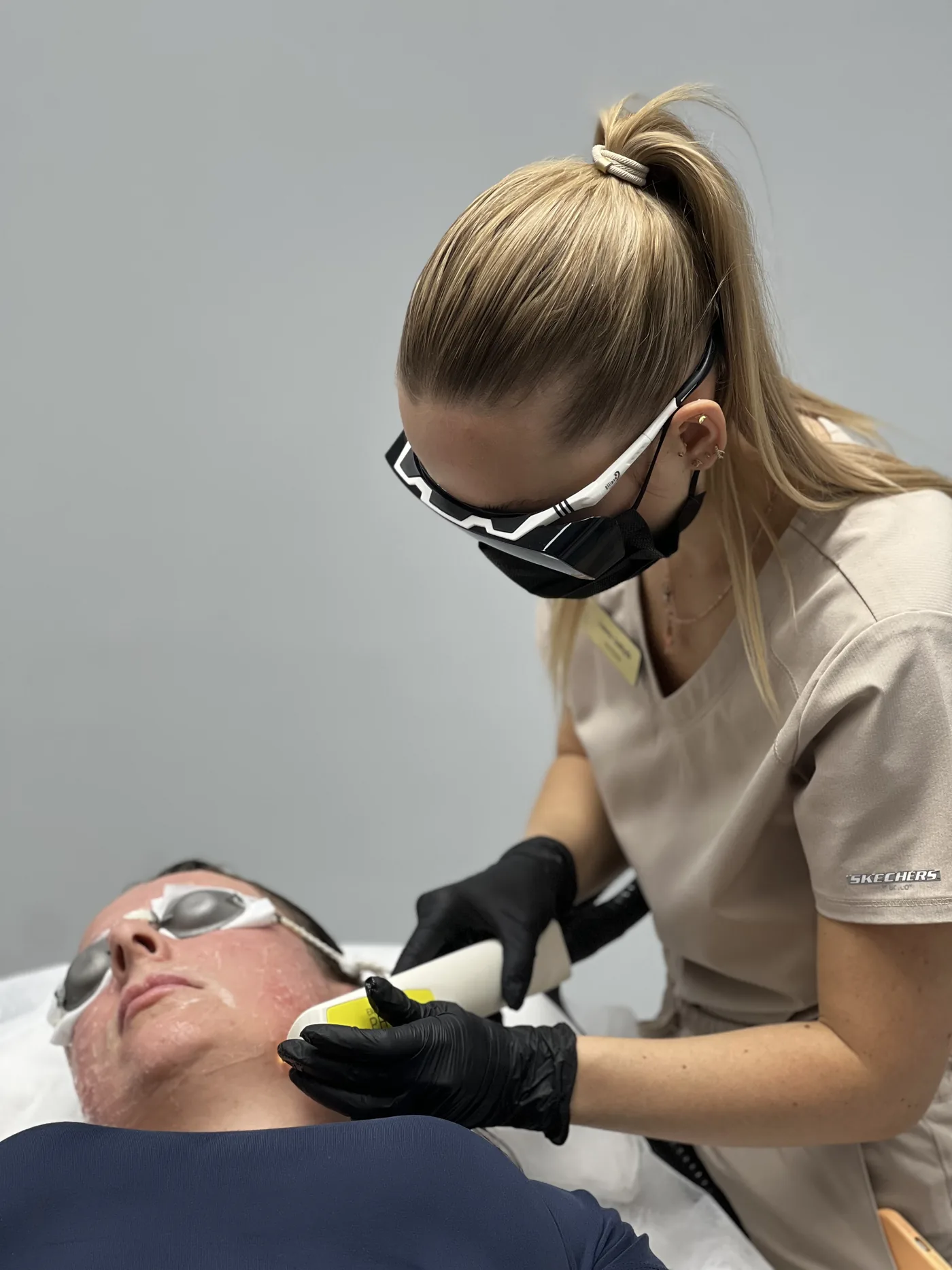
Pulses are delivered across the lesion, with skin cooling and monitoring of the reaction. Protective glasses are mandatory. We do not usually use anaesthetic, because it limits the constriction of the vessels.
4
Resorption and assessment
The lesion may temporarily darken before the body absorbs the closed vessels. The effect is assessed once resorption is complete, usually after a few weeks.
The body removes a closed vessel over a period of weeks, so the effect is assessed once resorption is complete, not in the week after the treatment. Aftercare is the same as for vessel removal, and we describe it on the treatment page, together with the recommendation that affects the result: limiting exertion that raises blood pressure.
What we do not do
We do not diagnose vascular lesions from a photograph or a description given over the phone.
We do not remove atypical, rapidly growing, ulcerated or bleeding lesions without prior diagnostics.
We do not treat port-wine stains or vascular malformations. This therapy is not yet part of our offering; we refer patients to centres that manage them systematically.
We do not treat varicose veins with a diameter larger than the method covers, or vascular insufficiency; this is the domain of vascular surgery and phlebology.
We do not treat vascular lesions in children.
This list is not a legal disclaimer, but a description of where the sensible scope of an aesthetic medicine practice ends. For lesions beyond it, we refer patients elsewhere, and we do so before the treatment, not after it.
Contraindications
A fresh tan postpones the treatment rather than ruling it out, but for the 532 nm range it is an absolute contraindication: the melanin in the epidermis then competes with haemoglobin for energy. This is why autumn and winter are a more convenient time for therapy of vascular lesions.
Editorial responsibility
Responsible for the content
NL Clinic, Katowice
How the content is created
Last updated
15 August 2026
This material is informational in nature and does not replace a consultation or qualification.
The questions we are asked most often
How does an angioma differ from a vessel?
A vessel is a single blood vessel that has permanently dilated, visible as a fine line. An angioma is a structure made up of overgrown vessels, with volume: raised or flat, but occupying space within the skin. The difference has a practical consequence, because an angioma usually requires more energy and, more often, several sessions.
Are cherry angiomas dangerous?
They are benign and very common lesions that appear with age. They are removed for aesthetic reasons. However, a lesion that grows quickly, changes shape, ulcerates or bleeds requires a medical assessment before any treatment, because not every red lesion on the skin is an angioma.
I have a port-wine stain, can you treat it?
No, this therapy is not yet part of our offering. A port-wine stain is a developmental vascular defect present from birth and requires management at a centre that treats such lesions systematically, often for years. At a consultation, however, we can help you recognise whether your lesion belongs to this group and point you in the right direction.
How many treatments will be needed?
It depends on the type of lesion, its volume and its depth. A single cherry angioma often clears after one treatment, while larger angiomas and malformations require a series. We determine the number of sessions after assessing the lesion, because the variation within this group is large, and giving a single figure would be misleading.
Does the treatment hurt?
Sensations are described as a burning feeling or brief stinging. We do not usually use anaesthetic, because numbing cream limits the constriction of the vessels, which is the reaction the patient comes for. Comfort is provided by cooling the skin before and after the treatment. For larger lesions and in sensitive areas, the approach is decided individually.
Why does the lesion look worse after the treatment?
This is usually the expected course, not a complication. Closed vessels may temporarily darken before the body absorbs them, and redness and swelling appear around the lesion. Resorption takes weeks, so the effect is assessed once it is complete, not in the week after the treatment.
Are vessels on the legs removed the same way as on the face?
No. Vessels on the legs lie deeper and have a larger diameter, so they require a longer wavelength, higher energy and a treatment carried out by a doctor. For lesions wider than the method covers, or if vascular insufficiency is suspected, the right direction is a phlebologist, not an aesthetic practice.
When is the best time to plan the treatment?
In autumn and winter, because a fresh tan is a contraindication, and the skin needs to be protected with sunscreen after the treatment. For the 532 nm range this is decisive, because the melanin in the epidermis then competes with haemoglobin for energy.
Where does poikiloderma on the neck and chest come from?
From chronic sun exposure in an area that is rarely protected with sunscreen. The lesion has two elements: vascular and pigmentary, so it requires work on both, not only on the redness. Without consistent photoprotection after therapy, the lesion recurs.
The content is for information. It does not replace a consultation, an examination or an individual assessment.