Important before booking
We do not remove pigmented lesions, that is moles and melanocytic naevi. Ablative methods vaporise the tissue, so no material remains for histopathological examination, and without it the nature of the lesion cannot be confirmed.
If the lesion is pigmented, has changed in appearance, colour, shape or size, itches, bleeds or worries you, the right path is a visit to a dermatologist with a dermoscopic examination. Removal of such a lesion, if indicated, is performed surgically, with histopathological examination.
The content is for information. It does not replace a consultation, an examination or an individual medical assessment.
On this page
What we remove and what we do not
This distinction matters more than the technique itself, which is why we put it first.
We remove
Milia, that is cysts filled with keratin.
Fibromas, including pedunculated ones, found in skin folds.
Seborrhoeic keratoses, after a doctor has confirmed their nature.
We refer to a dermatologist
Melanocytic naevi and moles, that is all pigmented lesions.
Lesions that have changed colour, shape, size or edges.
Lesions that itch, bleed, weep or ulcerate.
Lesions of unclear nature that raise the doctor's doubts during qualification.
We support the assessment of lesions with digital dermoscopy, which allows the lesion to be viewed under magnification and its appearance documented. It is decision support, not a diagnosis. In difficult cases we refer you to cooperating dermatologists rather than proceeding with a treatment on our own.
Every treatment is preceded by a doctor's assessment of the lesion. If during qualification any doubt arises about the nature of the lesion, we withdraw from the treatment and refer you for diagnostics, even if you came with a decision already made.
Milia
Milia are small cysts filled with keratin, lying just under the surface of the skin. They look like hard, white bumps resembling pimples, but they are not inflammatory lesions and cannot be squeezed out.
They usually appear in numbers, most often around the eyes, including on the eyelids, on the temples, forehead and nose, in people of all ages. They pose no risk to health and are not contagious, so the only reason to remove them is usually appearance or discomfort.
Fibromas
Fibromas, also called fibroma, are benign nodules built of collagen fibres and blood vessels, surrounded by a thicker layer of epidermis. They grow slowly and are skin-coloured or brown.
They occur most often in skin folds: on the neck, under the arms and in the groin, in women and in men. The problem is often not only appearance but also catching on clothing and jewellery, which leads to irritation.
The exact cause of their formation is not known. Contributing factors include frequent chafing and irritation where skin folds meet, elevated growth hormone levels, human papillomavirus and insulin resistance. People with obesity and type 2 diabetes are more at risk.
Seborrhoeic keratoses
These are benign lesions found in most people over the age of sixty, although they also appear earlier. Most often on the trunk, upper limbs, face, including the eyelids, and on the scalp. They do not hurt, itch or sting.
Despite the name they have nothing to do with the sebaceous glands or sebum. Their number grows with age, and contributing factors include sunburn and skin inflammation, friction, especially in folds, the presence of lentigines, hereditary predisposition and the use of epidermal growth factor receptor inhibitors.
They can be flat or raised and are well demarcated from healthy skin.
Diameter from 1 mm to several centimetres.
Colour yellow, grey, light brown, dark brown, black or multicoloured.
Surface smooth, waxy or warty.
They occur singly or in groups: on the scalp, under the bust, along the spine, in the groin.
These features help you get an idea of what you may be dealing with, but they do not replace a diagnosis. A seborrhoeic keratosis can be misleadingly similar to other lesions, which is why it is assessed by a doctor, and in case of doubt by a dermatologist with a dermatoscope.
CO2 laser and radiosurgical diathermy
Both methods work by vaporising the tissue of the lesion; they differ in the energy carrier. The choice belongs to the doctor and depends on the type of lesion, its size and location.
CO2 laser
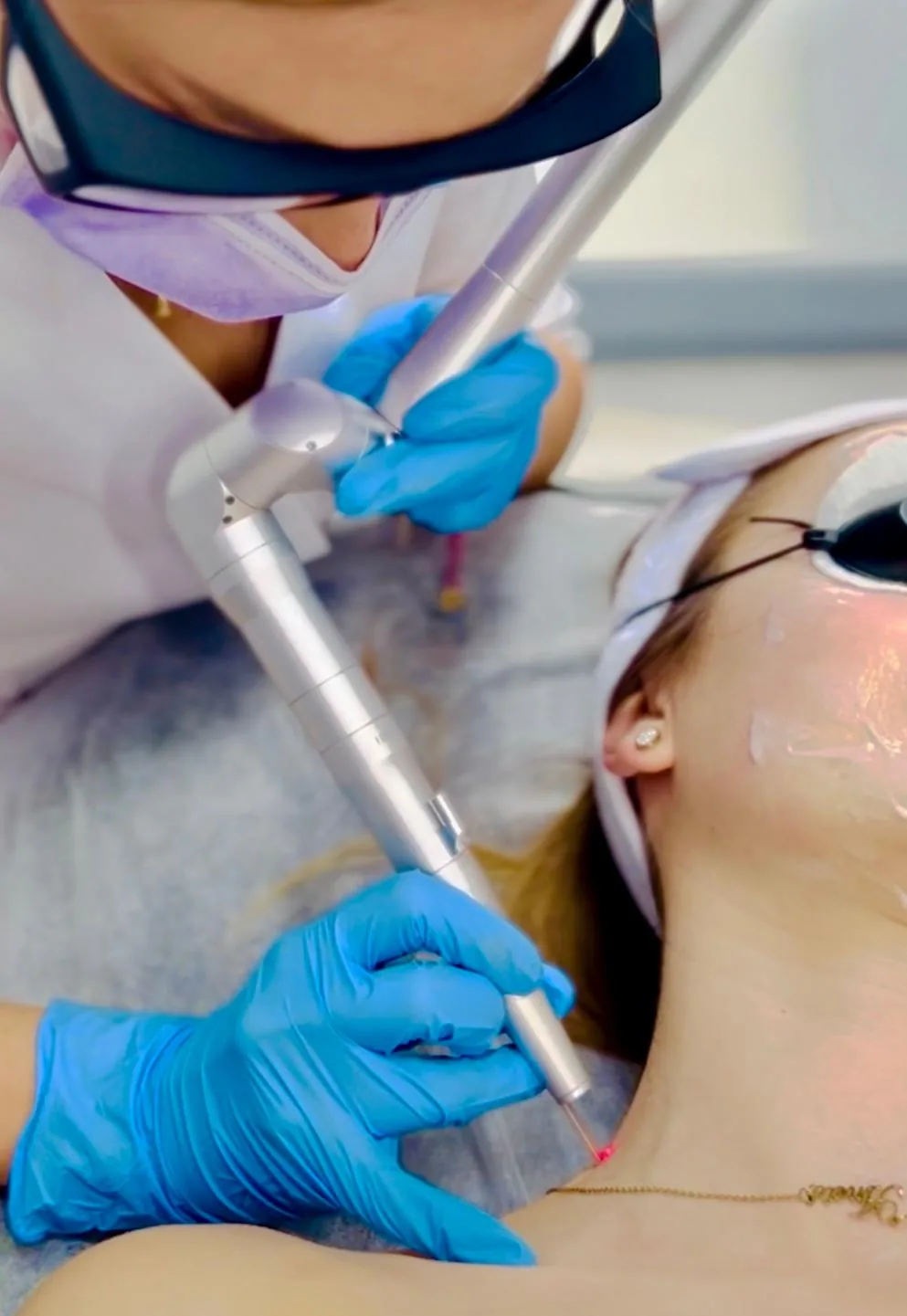
The beam vaporises the tissue of the lesion layer by layer, with great precision and depth control. It works well for small lesions and in areas requiring accuracy, for example on the eyelids.
Radiosurgical diathermy
High-frequency radio waves cut and coagulate the tissue at the same time. It is often chosen for larger and pedunculated lesions, where the vessels need to be sealed at the same time.
The treatment on video
The video will show how precisely the laser works and how big the tiny scab forming at the site of the lesion is. It answers the most common concern with this treatment, the question about a scar.
We do not publish before and after material. With skin lesions such comparisons are common in the industry and at the same time particularly misleading: the appearance of the site after the treatment is largely decided by healing and by whether the scab fell off on its own.
The treatment step by step
The treatment takes from ten to sixty minutes, depending on the number and size of the lesions. We use anaesthesia for comfort, as a cream or local anaesthesia, depending on the size of the lesion and your pain threshold.
Healing and aftercare
At the site of the removed lesion a tiny scab forms, which heals over three to seven days. Once it falls off, new skin is revealed. The treatment requires no recovery time, so you return to your daily activities the same day.
With milia and fibromas usually no trace remains. With larger seborrhoeic keratoses, especially pedunculated ones, a pink spot the size of the original lesion may persist for some time.
Do not pick off the scab
The scab must fall off on its own. Picking it off early is the most common cause of a scar and pigmentation at that site.
Sun protection
A high-factor sunscreen on the treated area for several weeks after healing. Fresh skin pigments very easily.
Wound hygiene
Gentle cleansing with the preparation recommended after the treatment. No colour cosmetics on the site until the scab falls off.
No swimming pool or sauna
For the first few days skip the swimming pool, sauna and long baths, because they soften the scab and increase the risk of infection.
When to get in touch
With increasing pain, spreading redness, purulent discharge or fever, contact us without waiting for the check-up.
Contraindications
With a tendency to keloids, qualification is individual, especially for lesions on the chest, shoulders and back, where the risk is higher. Report previous keloids, even if they formed after other procedures or injuries.
The questions we are asked most often
Do you remove moles?
We do not remove pigmented lesions, including moles and melanocytic naevi. The laser and diathermy vaporise the tissue, so no material remains for histopathological examination, and without it there is no way to confirm what the lesion was. Such lesions are assessed by a dermatologist, and if they require removal, it is done surgically, with an examination.
How do I know whether my lesion qualifies for the treatment?
You do not have to know that; that is what qualification is for. Come to a consultation, the doctor will look at the lesion and say whether we remove it here or refer you to a dermatologist. If the lesion has changed in appearance or worries you, book a dermatologist straight away.
Will there be a scar after the treatment?
With milia and fibromas usually no trace remains. With larger seborrhoeic keratoses a pink spot the size of the original lesion may persist for some time. The risk of a scar rises if the scab is picked off and with a tendency to keloids.
How long does the skin take to heal?
The tiny scab stays for three to seven days and then falls off, revealing new skin. The treatment itself requires no recovery time, so you return to your activities the same day.
Can several lesions be removed during one visit?
Yes, and it is a common situation, especially with milia and fibromas, which occur in numbers. The number of lesions affects the length of the treatment, which can then reach an hour.
Does the treatment hurt?
We use anaesthesia: a cream for small lesions or local anaesthesia for larger ones. The choice also depends on your pain threshold, so mention it during qualification.
Will the lesion grow back?
A removed lesion does not grow back in the same place, but the tendency to develop them remains. With fibromas and seborrhoeic keratoses new lesions may appear over time in other places, because the cause lies in the characteristics of the skin, not in a particular nodule.
Can the treatment be done in summer?
It can, but it requires consistent sun protection after the treatment. Fresh skin after the scab falls off pigments very easily, so for lesions on exposed parts of the body autumn is a more convenient time.
Will I get the result of an examination of the lesion?
No, because with ablative methods the tissue is vaporised and there is no material to examine. That is precisely why we remove by this route only benign lesions of unambiguous nature, confirmed by the doctor during qualification.
How much does removing a skin lesion cost?
The cost depends on the number and size of the lesions and on the chosen method, which is why we keep one current price list for aesthetic medicine. You will learn the exact price after the lesions have been examined at the consultation.
The content is for information. It does not replace a consultation, an examination or an individual medical assessment.