Book straight away if
The mole is growing or changing shape, colour or thickness, and the change is progressing over weeks or months.
Bleeding, oozing, a scab or an erosion appears without any earlier injury.
The lesion itches, stings or hurts, and the symptom persists.
An inflamed border, pigmentation or small satellite spots appear around the mole.
A new skin lesion in an adult that is not healing, or a lesion clearly different from the others.
A lesion detected earlier requires a smaller treatment and gives a better prognosis. Delaying has no advantage here, and a visit that ends with the finding that nothing concerning is happening is not a wasted visit.
On this page
The ABCDE rule and the ugly duckling sign
ABCDE is a screening tool, commonly used in patient education. It helps to organise self-examination, but it does not make a diagnosis and does not detect every lesion that needs treatment. Some lesions do not meet any of these criteria.
A
Asymmetry
One half of the lesion does not match the other when you imagine an axis running through it. Benign lesions are usually symmetrical.
B
Border
Irregular, jagged, blurred borders or borders with projections. Benign moles usually have a smooth, well-defined border.
C
Colour
More than one shade within a single lesion: brown next to black, red, white or a bluish tone. A colour that changes over time is also concerning.
D
Diameter
Above about six millimetres, in other words larger than the top of a pencil. This is a supporting criterion: lesions that need treatment can be smaller, and large moles can be entirely benign.
E
Evolution
Any change over time: in size, shape, colour, thickness, surface. This criterion is the most important and at the same time the hardest to notice without photographs.
The ugly duckling sign
One person's moles are usually similar to each other. A lesion that clearly differs from the others, in colour, shape or size, deserves attention even when it does not meet the ABCDE criteria. This is a signal that is easier to notice at home than measuring the diameter.
Taking photographs helps: the same mole, in the same lighting, with a ruler or a coin for scale, every few months. Evolution is the hardest thing to notice from memory and the easiest to document.
What dermoscopy involves
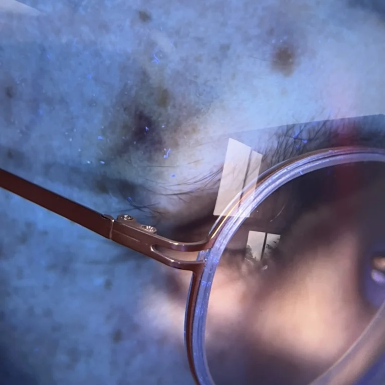
A dermatoscope is an optical device that examines the skin under magnification, in polarised light. It shows structures invisible to the naked eye: pigment pattern, network, vessels. The examination is non-invasive, painless and requires no preparation.
What dermatoscopy can determine, and what it cannot
It determines what to do next: observe, review after a set time, or remove the lesion and send it for examination. It also helps distinguish pigmented lesions from non-pigmented ones, which matters when qualifying for treatments.
It does not provide a histopathological diagnosis. The nature of a lesion is established only through microscopic examination of the removed tissue, not from an image, a photograph or an application.
Why moles are not removed with a laser
This question comes up often, usually on the assumption that laser is a gentler method leaving a smaller scar. The problem does not lie in the scar.
Laser vaporises tissue
After a laser treatment, no material remains that could be examined under a microscope. The lesion disappears, and so does the only way of establishing what it was. If it required treatment, the diagnosis is delayed, and the lesion may regrow in an altered form that is harder to assess.
This is why melanocytic naevi, that is ordinary moles, are removed surgically, with a margin, and sent for histopathological examination. This is standard practice, not excessive caution.
Laser is used, however, for some non-pigmented lesions, after a prior medical assessment ruling out any doubts. The scope of such treatments is described on the skin lesion removal page.
It is worth saying separately that removing skin lesions in practices without a medical assessment, for example during a cosmetic treatment, is a practice we keep our distance from. It is not about the technique, but about the omitted step that cannot be undone.
Not every skin lesion is a mole
Some lesions that patients call moles have a completely different origin. The distinction is for the doctor to make, but knowing that the categories differ helps explain why the approach can vary.
Melanocytic naevi
Ordinary moles, made up of pigment cells. They can be flat or raised, congenital or acquired. The rule of surgical removal with histopathological examination applies to these.
Seborrhoeic keratoses
Brown, rough, as if stuck onto the surface of the skin, appearing with age. They are benign and unrelated to pigment cells, but are sometimes mistaken for naevi.
Fibromas and pedunculated lesions
Soft growths, most often on the neck, under the arms and in the groin. Usually benign, often removed because they are irritated by clothing or jewellery.
Vascular lesions
Haemangiomas and small changes with a red or bluish colour, resulting from the structure of the vessels, not from pigment. They are described on a separate page about haemangiomas and vascular lesions.
There is one common rule: which category a particular lesion belongs to is decided by examination, not by resemblance to a photo on the internet.
Who should check their skin more often
The frequency of check-ups is set by the doctor individually. The following factors usually prompt more frequent observation, and their presence does not mean disease.
Fair complexion, red or light hair, skin that burns in the sun instead of tanning.
Sunburn in childhood and youth, especially with blistering.
A large number of naevi on the body, or naevi with an atypical appearance.
A history of skin cancer in the patient or in a first-degree relative.
Use of sunbeds, as well as work or activity in intense sun without protection.
Reduced immunity, including after organ transplantation and during immunosuppressive treatment.
Regardless of the scheduled dates, one overriding rule applies: a change noticed by the patient is a reason for an earlier visit, not for waiting for the next check-up.
Moles and laser treatments in another area
The mere presence of naevi does not rule out laser treatments or hair removal. It does, however, require a skin assessment before qualification and a few rules during the procedure.
We do not irradiate naevi. Lesions located within the treatment area are avoided and protected, because energy absorbed by melanin can change their appearance and make later assessment more difficult.
In laser hair removal, this also applies to areas densely covered with small naevi. In that case, the shielding is part of preparing the area, not an obstacle.
If a lesion of concern is present within the treatment area, the treatment in that area is postponed until a dermatological assessment has been carried out.
What this looks like at our practice
Observation from the practice
We examine the skin in the treatment area with a dermatoscope before every qualification for a laser treatment. This is not a full body screening examination, but it catches lesions that should not be irradiated, and it can be the moment when a patient learns of a lesion that needs assessment.
We do not provide oncological treatment. When a lesion raises concern, we do not carry out the treatment, and we say so directly, even when the patient came in for something completely different.
This is the practice's own approach, not data from the literature. We mark it separately so that it does not get mixed up with what comes from publications.
Editorial responsibility
Responsible for the content
NL Clinic, Katowice
How the content is created
Last updated
15 August 2026
This material is informational in nature and does not replace a consultation or qualification.
The questions we are asked most often
How can you tell that a mole needs a medical assessment?
The ABCDE rule is helpful: asymmetry of shape, irregular borders, uneven colour, a diameter of more than about six millimetres, and evolution, meaning a change over time. A separate signal is the ugly duckling sign: a mole that clearly stands out in appearance from the others on the same body. A medical assessment is also warranted for any bleeding, oozing, itching, pain, or a lesion that does not heal despite several weeks passing. The ABCDE rule is a screening tool, not a diagnosis, and it does not detect all lesions that require treatment.
Can a mole be removed with a laser?
Melanocytic naevi, that is ordinary moles, are not removed with a laser. Laser vaporises tissue, so no material remains that could be examined under a microscope, and this examination is the only way to establish the nature of a lesion. Removing a pigmented lesion without histopathological examination can delay the diagnosis of a disease requiring treatment. Laser is used, however, for some non-pigmented lesions, after a prior medical assessment.
What is a dermatoscopic examination?
It is the examination of a lesion under magnification, in polarised light, using a dermatoscope. It allows structures invisible to the naked eye to be seen and helps assess whether the lesion requires further action. The examination is non-invasive, painless and requires no preparation. It does not provide a histopathological diagnosis: it determines what to do next, not what the lesion is.
Do all moles need to be removed as a precaution?
There is no basis for the preventive removal of naevi that raise no concern on medical assessment. The treatment of choice is observation and follow-up, and removal is reserved for lesions of concern and those that are constantly irritated mechanically. Removing healthy naevi does not reduce the risk of disease, and it leaves scars.
Is a laser treatment in a different area safe if I have a lot of moles?
Pigmented lesions are not treated with light-based devices. During laser and IPL treatments, naevi within the treatment area are avoided and protected, because energy absorbed by melanin can change their appearance, which makes later assessment more difficult. The number of naevi alone does not rule out a treatment in another area, but it does require a skin assessment before qualification.
A naevus has been scratched and is bleeding. Is this a reason for concern?
A single mechanical injury, for example a scratch or rubbing from clothing, is not on its own a warning sign and does not turn a healthy naevus into something else. What does call for assessment is bleeding that appears without injury, recurs, or affects a lesion that has already changed. Naevi that are constantly irritated mechanically are also worth showing to a doctor for practical reasons.
How often should naevi be checked?
The frequency of checks is set individually by the doctor, taking into account the number and appearance of the naevi, skin phototype, past sunburn and skin cancer in the family. Regardless of the schedule, the rule is that any change noticed by the patient is a reason for an earlier visit, not for waiting for the appointed date.
Does NL Clinic carry out diagnostics of pigmented lesions?
We assess the skin with a dermatoscope before every qualification for a laser treatment, and this is when we pick up changes that need further action. We do not provide oncological treatment and we do not carry out treatments on lesions that raise doubt. In that situation we say so directly and refer the patient to a dermatologist.
The content is for information. It does not replace a consultation, an examination or an individual assessment.